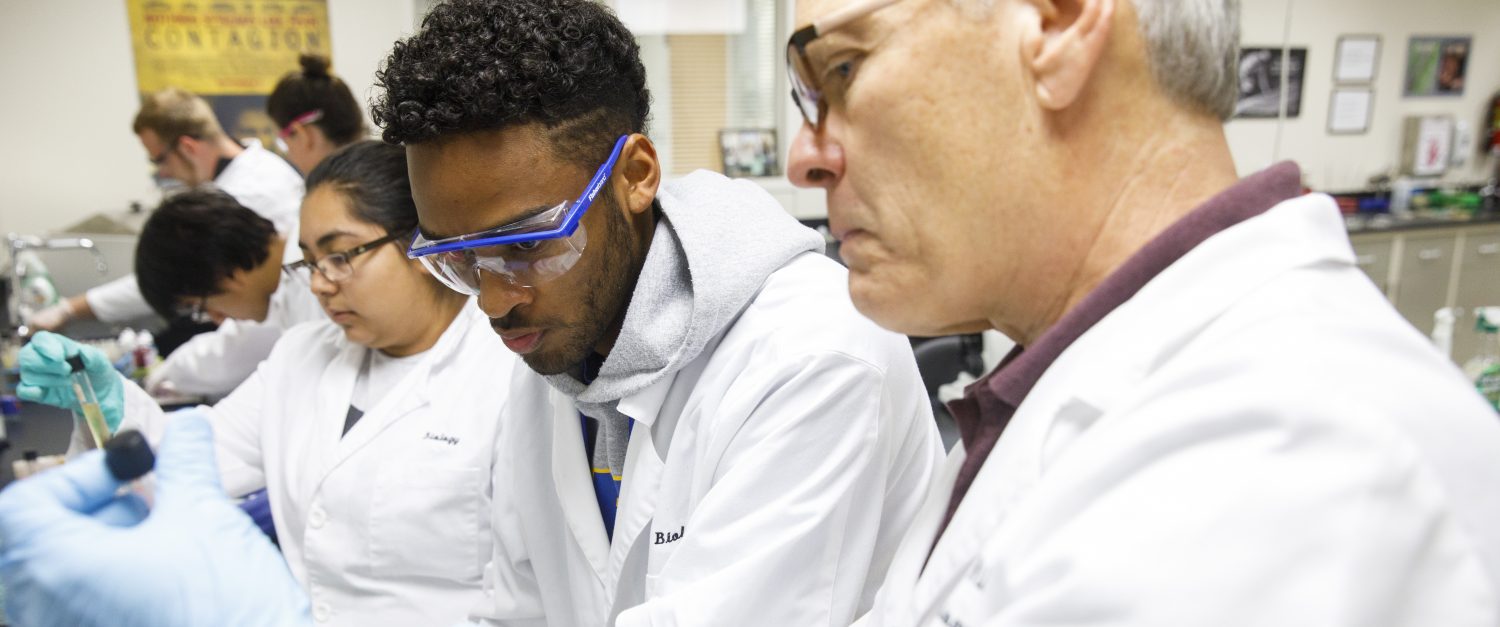


Today, McMurry science faculty and students had representatives from Nanoscience Instruments here to give us a hands-on demonstration of their benchtop scanning electron microscope – the Phenom XL.
Between 9 and 2, students and faculty to see the instrument in action. Once they participated in a demonstration for how to operate, everyone was able to bring or suggest samples and scan them. Once everyone learned how it worked, they were given the opportunity to use it. A microscope like this is versatile and useful for a variety of projects from materials science, physics, chemistry, and biology.
This particular microscope is easy to use and requires less sample preparation. Maximum magnification is 100,000x with an optical navigation camera using 3 – 16x magnification. Everyone was very impressed with the easy to use software and left with quite a few ideas about how the SEM could extend what they are doing in class and with research teams.

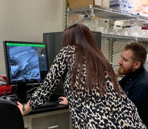
Dr. Zach Gray demonstrates the Phenom XL 
Unusual feature from collagen studies 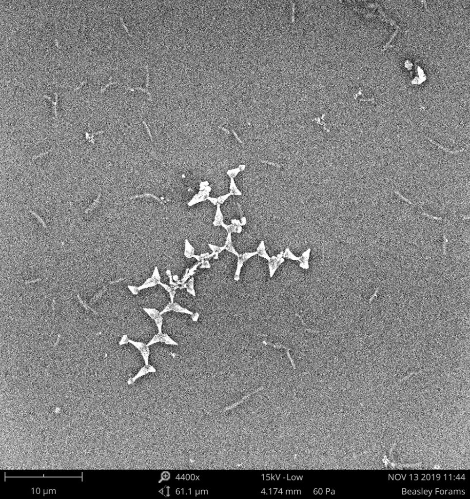
Another unusual feature from Team Oncogeniuses 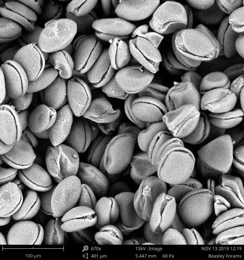
Dr. Anna brought in some monocot pollen. 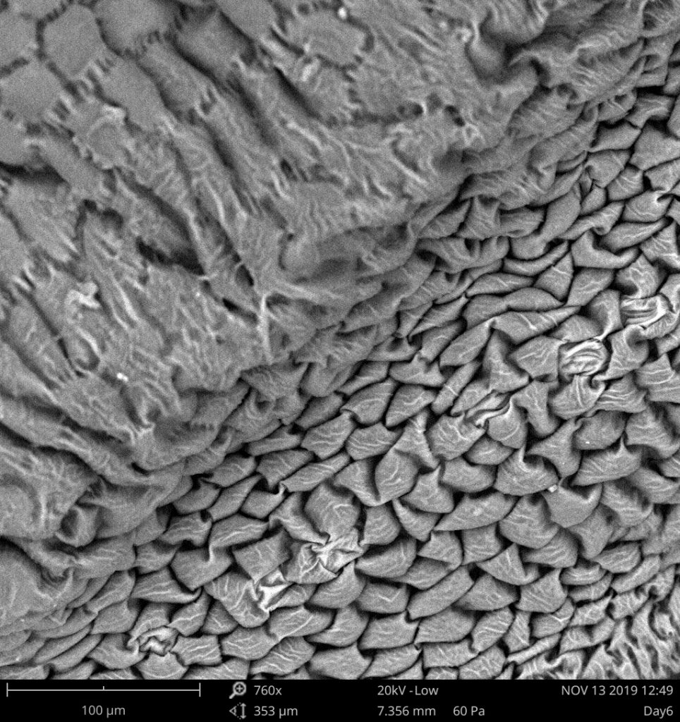
Any guesses? 
Pollen on plant tissue. 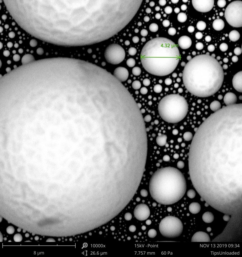
Part of the demo was looking at some standard materials – metallic particles. 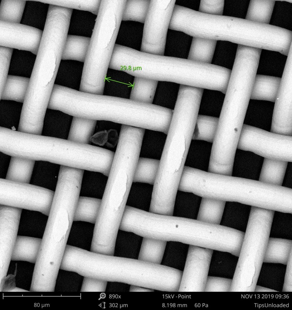
A wire mesh with grid size below detection by the eye. 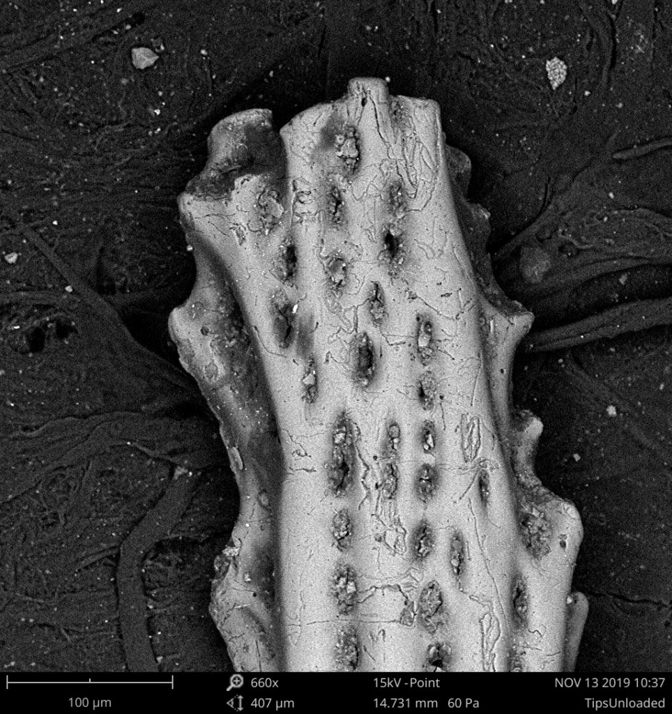
End of a calcified specimen from the archived collection left by Dr. Beasley.