
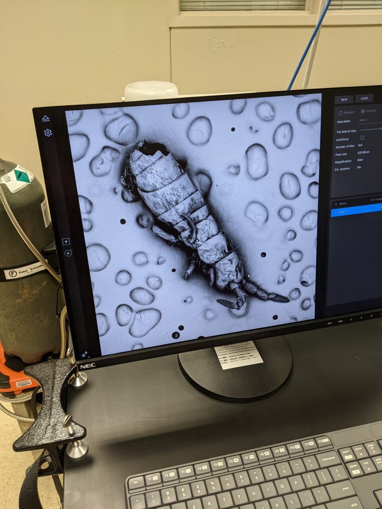
Here’s the whole springtail. Typically about 1 mm long. 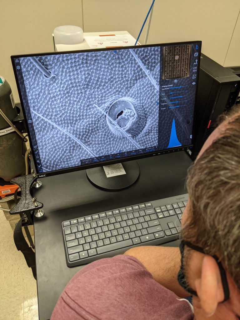
The images blew us away. 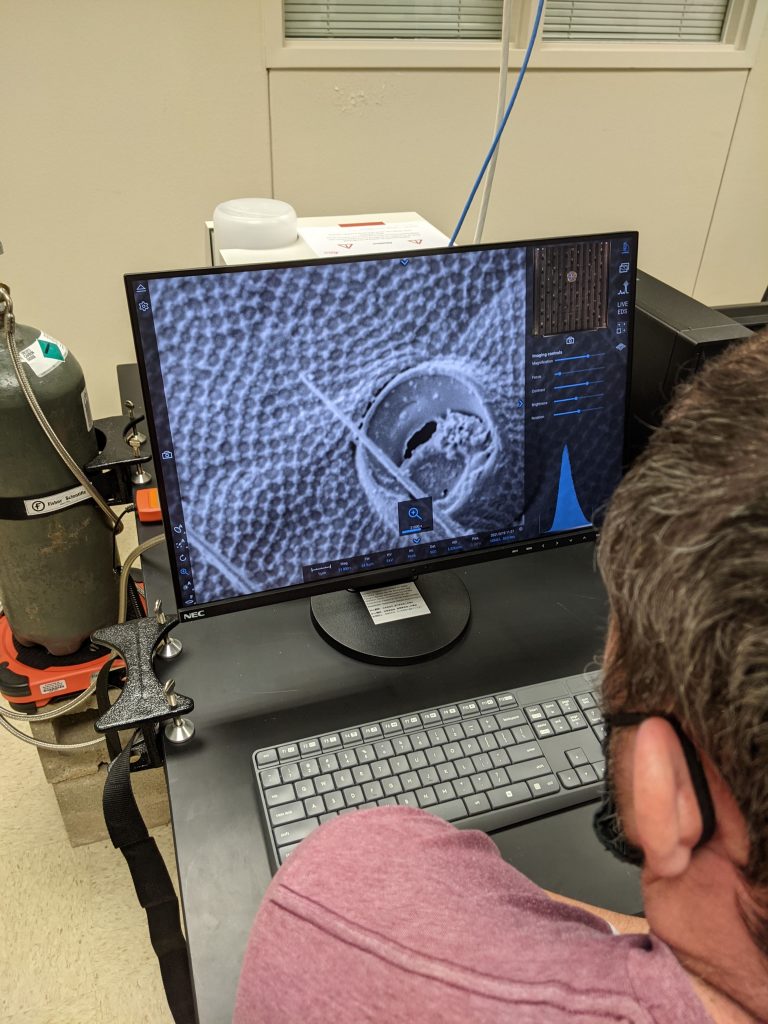
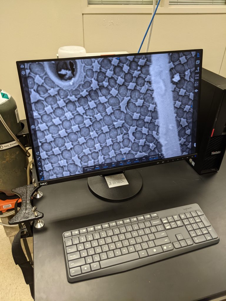
Repetitive pattern of proteins making exoskeleton 
Not bad for 152,000x 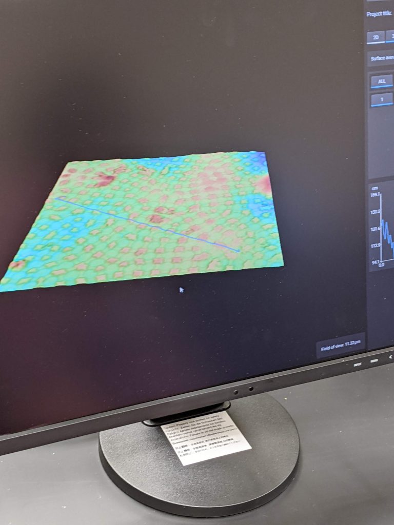
By changing illumination, you can create 3D images that can be fed into a 3D printer to create a model of the insect exoskeleton surface. 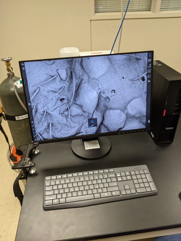
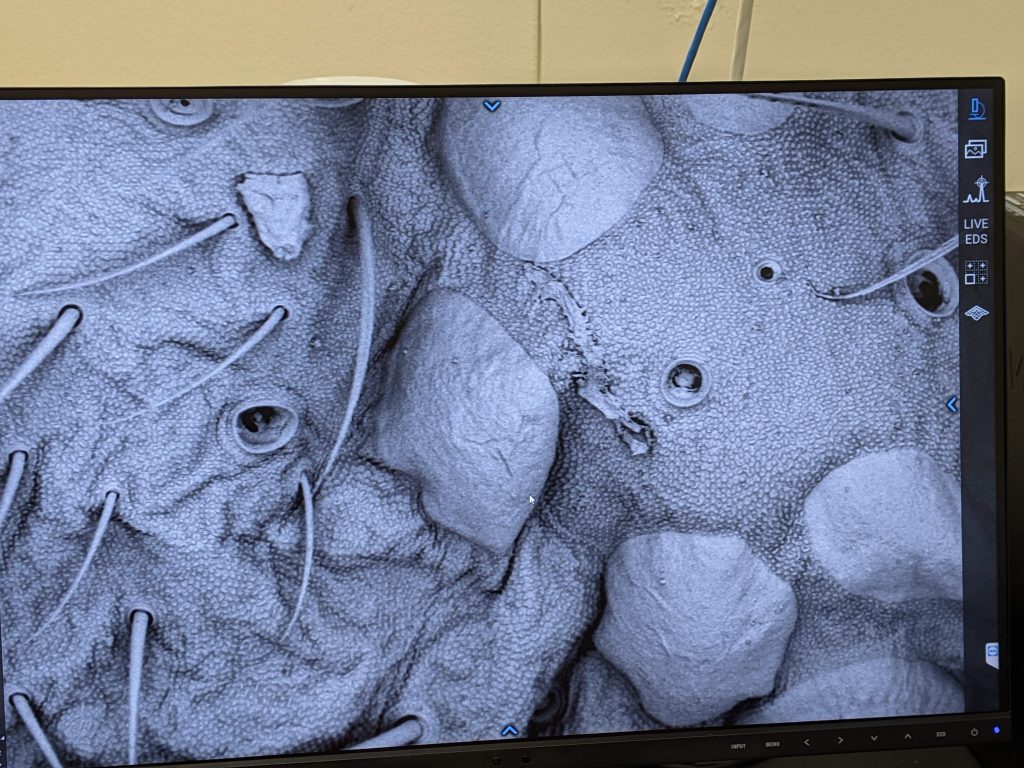
Structures near the mouth parts of the springtail. 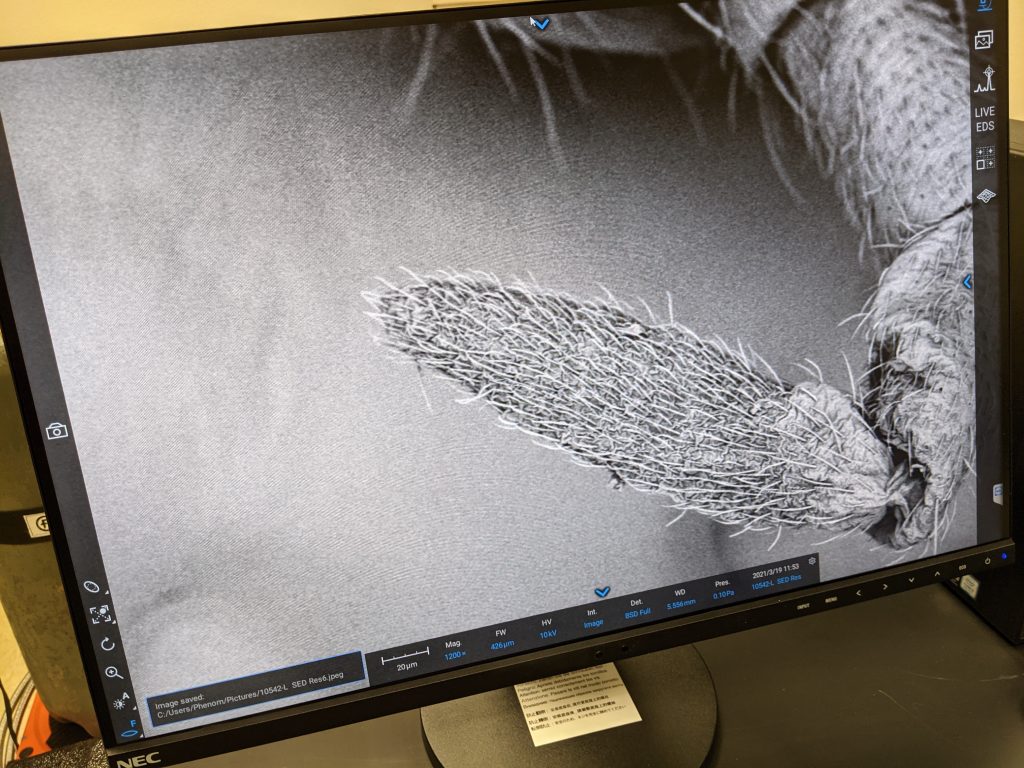
Fine detail of the end of an antenna of the springtail. 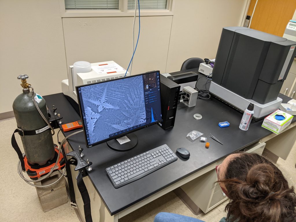
Dr. O’Connell viewing collagen and cancer cells (but mostly residual salt crystals). This was an attempt to see structures from a sample not created for SEM viewing.
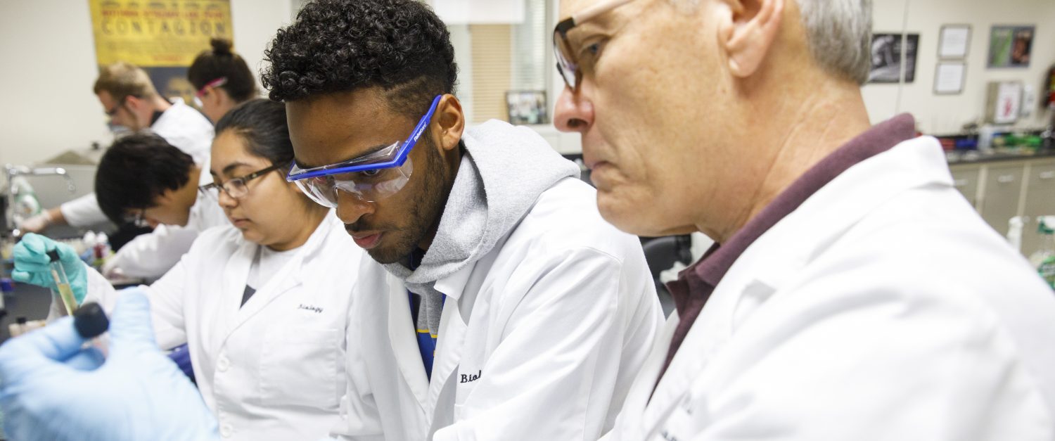
THANK YOU TITLE V PARTNERING FOR STEM SUCCESS FOR PROVIDING SUCH A GREAT ADDITION TO OUR RESOURCES FOR RESEARCH TEAMS!