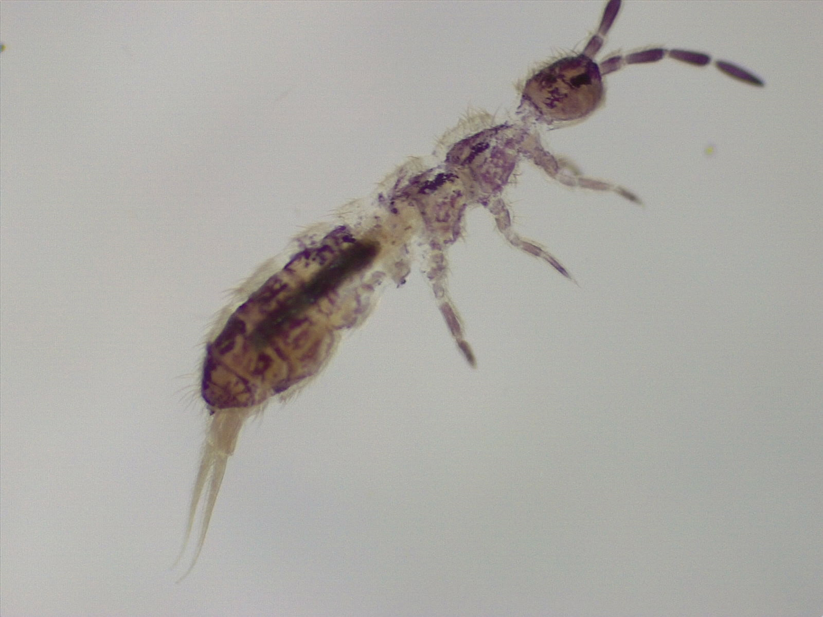
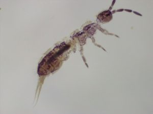
WOW!! WOW!! WOW!! That’s all you can say when you run your first scan using an SEM. This semester finds me teaching an Entomology course so I chose a small insect like creature called a springtail as the first specimen to look at. The first picture below is what a springtail looks like using a dissecting scope.
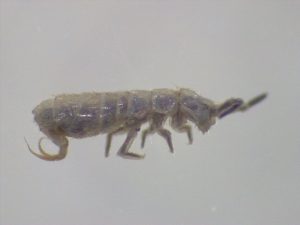
In order to visualize the specimen using the SEM I needed to prep the sample by running it through the new Critical Point Dryer. This machine replaces the fluid inside the specimen with liquid CO2 that evaporates when heated leaving the specimen dry. The next image shows the springtail after drying.
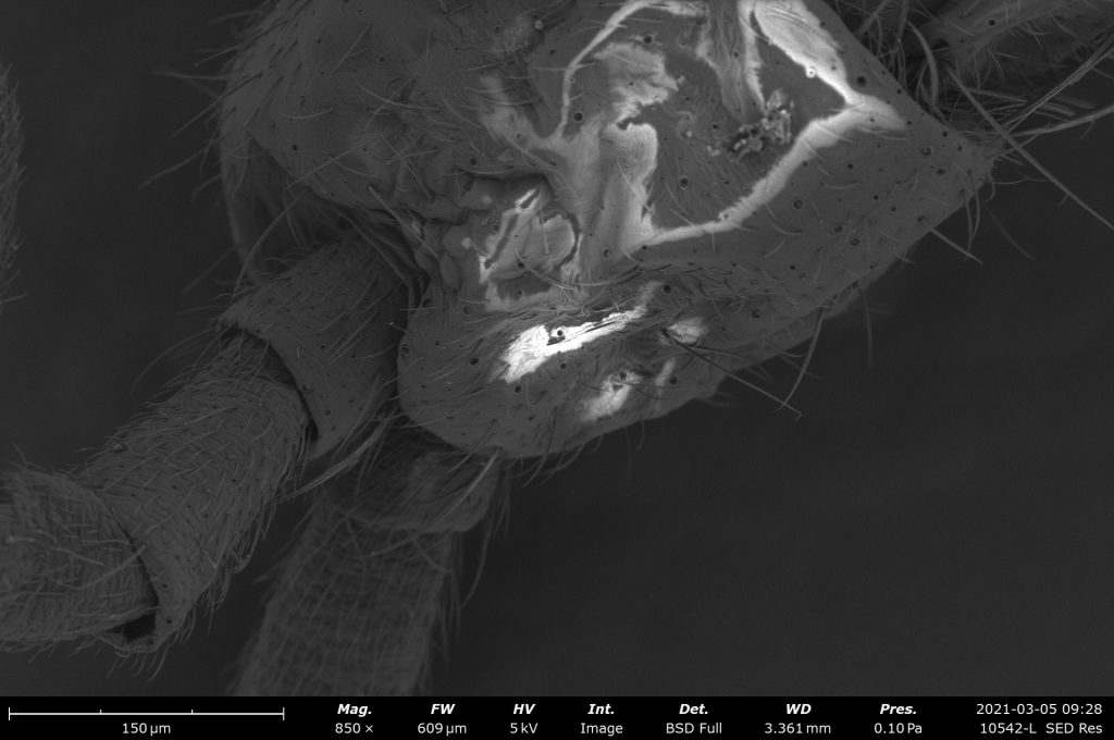
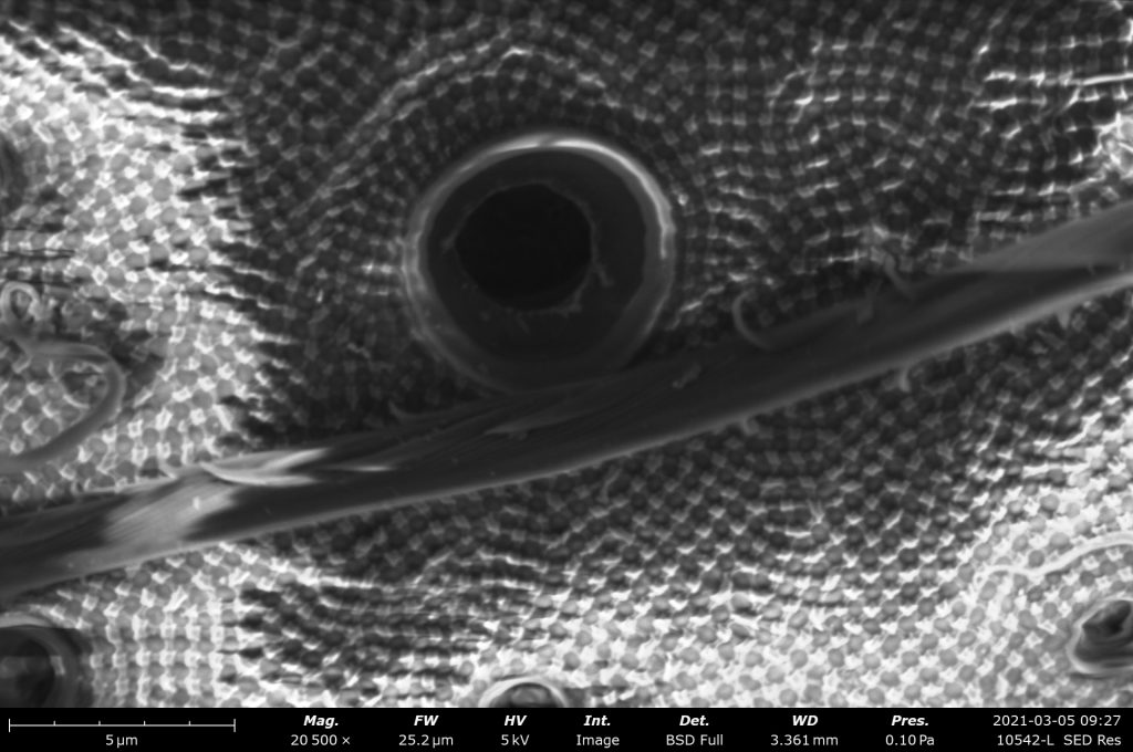
Now for the really cool stuff. I made the first run with the new Phenom Scanning Electron Microscope this morning. The following images highlight three different areas. The first shows the head and the base of the antennae at 850x. The second shows the lower segments (called tarsi) of the front leg at 1700x. The final image is zoomed in to a whopping 20,500x showing a close up of the chitin exoskeleton. You can see the a hair running left to right and actually see the individual protein fibers that compose it! What’s really amazing is the machine will go up to just over 200,000x total magnification! This piece of equipment will certainly usher in a new aspect to all of the awesome research going on here at McMurry!