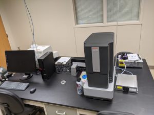
 It is hard to believe the investment in this room! You are looking at a new generation Phenom XL scanning electron microscope capable of over 200,000x magnification. Besides allowing us to see biological specimens as never before, it has the ability to conduct elemental analysis (what and the percentages) and to perform tensile strength analysis for materials science. To support the use of this instrument, we also have a new automated Leica CPD 300 critical point dryer and a Luxor carbon/gold sputter coater (putting a thin layer of a conducting material on the surface enhances secondary electrons for a detailed image).
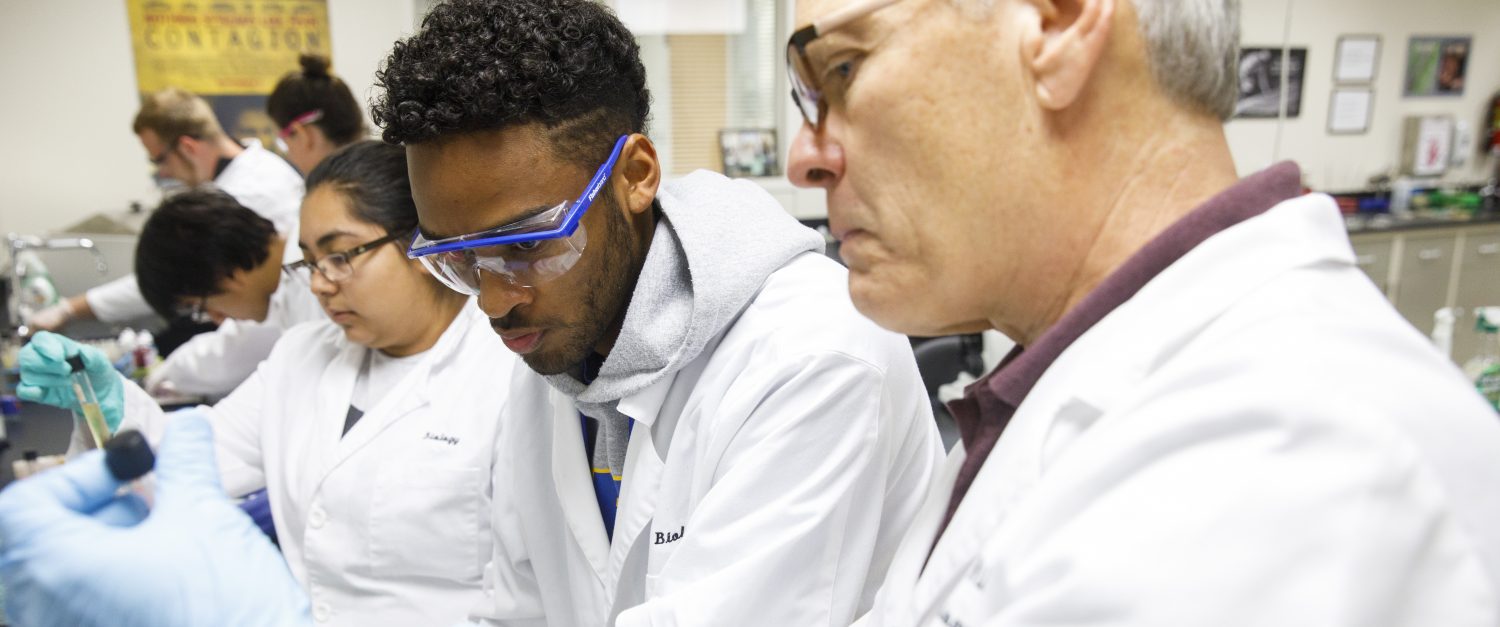
Dr. Boyle was the first in line to use the instrument. He performed critical point drying on an invertebrate called a springtail yesterday and was able to observe the specimen at over 4,000x this morning. The detail was STUNNING! And that was without sputter coating! Wow! (We’ll be saying that a lot from now on!)
Over the next couple of weeks we will find opportunities for core faculty from our campus to train on the use of these new pieces of equipment. Our expectation is that a majority of our Research Teams will feature SEM images in their research presentations this April!
And this is not just a McMurry thing! The instrument can be operated remotely, allowing our Cisco partners the opportunity to benefit. Faculty can bring their materials to campus, prepare them for viewing and do their microscopy and capture their images. They can even operate the microscope from their own campuses…we load, and they access the Phenom remotely and capture the images they want. All it takes is a computer and the Internet.
We expect to host workshops this summer to train faculty from across our campuses on how to prepare and view and capture images with this instrument. Wow – a new era is beginning for our campus and friends thanks to Title V!